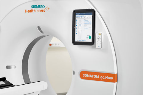
Computer Tomography (CT) is based on the use of X-ray. The domain of CT diagnostics are acute changes in the brain, emergency and trauma diagnostic as well as diseases of the chest and abdomen, vessels and bones. In addition to established primary use of CT, it is also possible for the diagnostic of tumours in the chest or abdomen. CT is in many cases both alternative and additional examination tool to better narrow a diagnosis.
Preparation
In certain cases it is necessary that you must be fasting for a few hours before the examination, for example to assess the gallbladder. In this regard, you will be informed when you become your appointment.
- Please check your medication and inform us if you are taking any medications for the thyroid or diabetes.
- Inform us when you already have been operated in the study area.
- If you have one, please bring your allergy passport with you.
- When you had a previous examination of the study area, please bring the examination (for example a CD) with you to your appointment.
Procedure
You will be asked to fill out the questionnaire, which you will find below or you will become at our reception. Please read the questionnaire carefully. The radiographer will come and get you and will discuss your questionnaire with you. You’ll be asked to change into a gown and remove any metal objects. Your personal items can be stocked in the changing room, which can be locked. You have to lie on a table, that slides into the CT scanner. The radiographer leaves the room during the examination and he/she goes into the control room where he/she can see and hear you. The radiographer will inform you about the examination, the duration will be between 10 and 20 minutes. For the examination of the chest and abdomen you will hear breathing commands (to hold your breath), to prevent your chest from moving up and down. The most import thing is that you lie still while the CT images are being taken.
An CT examination of the kidneys takes a little bit longer, between 20 to 30 minutes.
The use of oral and/or rectal contrast material
To visualize the gastrointestinal tract and the abdomen, it is often useful to drink the contrast material or to administer the contrast material in the rectum. When oral contrast material is necessary, you need to come an hour before the real examination. It’s important to drink the contrast material evenly over an hour, for better examination quality. The stomach and intestines transport the contrast material forward. If rectal contrast material is necessary, you don’t need a special preparation and it’s performed shortly before the examination starts. The rectal contrast material can make you feel bloated an uncomfortable. The use of oral and rectal contrast material, makes it easier to show changes in the intestine and the intestine can be better differentiated from other anatomical structures.
Side effects: Rarely it can lead to slight diarrhea.
The use of intravenously contrast material
Intravenously contrast material is used to show the parenchyma of the organs and blood vessels. The contrast material is an Iodine-containing substance and very important for precise diagnostics. The use of intravenously contrast material will be checked by the radiologist. You will be informed and prepared by the radiographer. You may experience a feeling of warmth during the injection, metallic taste in your mouth and/or the feeling you have to pee. These feelings are normal and harmless. There are no restrictions after intravenously contrast material, you can drive a vehicle afterwards or go back to your work. We recommend you to drink a lot of water, because the contrast material is being excreted through the kidneys and urine. Important: Please inform the radiographer when you’re pregnant, you have diabetes, hyperthyroidism or a kidney disease.
Contrast material allergy
Please inform your doctor and the radiographer when you previously had an allergic reaction to the contrast material. The examination will be performed without contrast material or only after previously injected anti-allergic drug and steroids. This counteract any potential side effect. Side effects: Today’s contrast material is very well tolerated.
In very rare cases it may cause itching, skin rash, sneezing and problems with swallowing. The risk of a severe allergic reaction is about 1:100’000. For each case we have the appropriate emergency medicines ready for use.
Radiation
As already mentioned above, during a CT scan you are briefly exposed to X-ray. Before each examination, the medical benefit of a CT scan is checked by the radiologist. At the same time, the radiation is adjusted to the appropriate examination and your physical condition. Different techniques and calculation algorithms allow us to perform the examination with minimized radiation. The radiation from our CT examinations are verified, evaluated and reviewed by the Federal Office of Public Health (Bundesamt für Gesundheit(BAG)).
Questionnaire
| German |
English |
Italian |
| Albanian |
Portuguese |
