Magnetic Resonance Imaging (MRI) is the central pillar of modern image diagnostics. In particular, diseases of the brain and spinal cord, as well as diseases of the bones, joints, spine and blood vessels are frequently examined by MRI.
But also special indications, for example the pelvis and abdomen, female breast and prostate, the MRI provides excellent diagnostics.
MRI Examination
When you had a previous examination of the study area, please bring the examination with you to your appointment (for example a CD).
Procedure
You will be asked to fill out the questionnaire, which you will find below or you will become at our reception. The radiographer will come and get you and will discuss your questionnaire with you. Please inform us when you have pacemaker, any other metal objects/medical devices in your body or when you’re pregnant. You’ll be asked to change into a gown and remove any metal objects. As the MRI produces strong magnetic fields, your personal belongings (phone, cash cards, jewellery) can be damaged. Your personal items can be stocked in the changing room, which can be locked. For some examination contrast material intravenously is necessary. This makes certain tissues and blood vessels show up more clearly and in greater detail. The radiographer will inform you. You will lie on a motorised bed, that moves inside the scanner. During the examination you’ll become an emergency button and the radiographer explains you when and how to use it wisely. If you use the button, be aware that the scan needs to be stopped and the radiographer comes to you.
Unfortunately, the scan has to be repeated. The MRI will make loud tapping noises during the examination, therefore you become headphones and it’s possible to listen music. Sometimes there won’t be any tapping noises, that’s normal. Important for the examination is that the body part, that is being examined, is in the middle of the scanner so that the examination is of the best possible quality. To avoid the images being blurred, it’s very important that the body part is still throughout the whole examination until the radiographer moves you out of the scanner. During the examination the radiographer leaves the room and will watch you from the outside. Your MRI scan will be studied by our radiologist and will send the report to your doctor (who arranged the scan) and will discuss the results with you at your next appointment.
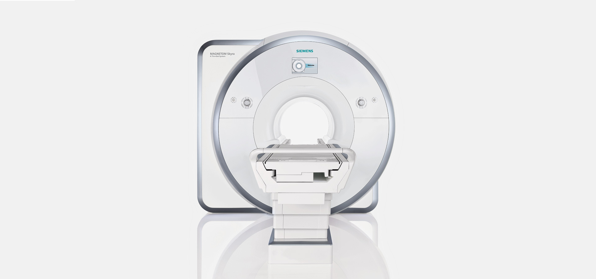
Claustrophobic
If you’re claustrophobic, please inform us when you make the appointment with the receptionist. When preparation is necessary, such as a drug, we need to adjust the appointment. It is also possible to arrange an appointment to take a look at the MRI scanner before we plan an MRI. In most cases an MRI can be performed without any drugs, the tube of the scanner is lit and the MRI ‘Skyra’ has a diameter of 70 cm. Often it helps when the eyes are closed during the MRI examination, because this reduces the narrow feeling. If you still need drugs, please bring someone with you so we can dismiss you with clear conscience. You are given a light sedative and you’re not allowed to drive a vehicle afterwards.
The use of contrast material
For many examinations it’s necessary to inject contrast material intravenously. The contrast material is a Gadolinium-containing substance and very important for good diagnostics. The use of contrast material intravenously will be checked by the radiologist. You will be informed and prepared by the radiographer. Normally the contrast material shouldn’t be noticed, but any warmth in the body is normal and harmless. Side effects: Today’s contrast material is very well tolerated. In very rare cases it may cause itching, skin rash, sneezing and problems with swallowing. The risk of a severe allergic reaction is about 1:100’000. For each case we have the appropriate emergency medicines ready for use. There are no restrictions after intravenously contrast material, you can drive a vehicle afterwards or go back to your work. We recommend you to drink a lot of water, because the contrast material is being excreted through the kidneys and urine. Important: Please inform the radiographer when you’re pregnant or when you have a kidney disease.
Contrast material allergy
Please inform your doctor and the radiographer when you previously had an allergic reaction to the contrast material. The examination will either be performed without contrast material or only after previously injected anti-allergic drug and steroids. This counteract any potential side effect.
Questionnaire
| German |
English |
Italian |
| Albanian |
Portuguese |